X-ray spectromicroscopes combine the advantages of x-ray spectroscopy and x-ray microscopy in a single device. X-ray absorption spectroscopy is sensitive to elemental composition and oxidation state, and requires a source of tunable x-rays – synchrotron radiation. There a number of way of imaging with x-rays, and performing laterally resolved spectroscopy.
The SPHINX and MEPHISTO instruments used by our group are X-ray PhotoElectron Emission spectroMicroscopes – X-PEEM. SPHINX (Spectromicroscope for PHotoelectron Imaging of Nanostructures with X-rays) Elmitec PEEM III and MEPHISTO (Microscope a Emission de PHoto_lectron par Illumination Synchrotronique de Type Onduleur).
The X-PEEM forms magnified images of a specimen surface by collecting and focussing photoelectrons. The field of electron imaging with electrostatic lenses was pioneered by Gert Rempfer (UV-PEEM), Brian Tonner (X-PEEM) and Ernst Bauer (LEEM) The lateral resolution does not reach that of electron microscopy because of lensing aberrations, but is presently better than 20 nm for MEPHISTO and 6 nm for SPHINX.
The spectromicroscopes use electrostatic or magnetic lenses to form a photoelectron image at the detector (see links above for specifics). The sample is held at a high negative voltage, while the first element is grounded, resulting in a strongly accelerating electric field between the sample and the optics. The detector is a multichannel plate that amplifies the electron signal, followed by a phosphor screen that provides the light image that is recorded by a video camera.
Note that there is no energy filtering of the photoelectrons – the total electron yield of electrons is captured by the X-PEEM. The electron yield is proportional to the x-ray absorption cross section. The transmission of the optics is most efficient for the low energy (1-5 eV) secondary electrons that result from inelastic scattering of primary and Auger electrons within a surface layer of 50 – 100 Å.
MEPHISTO
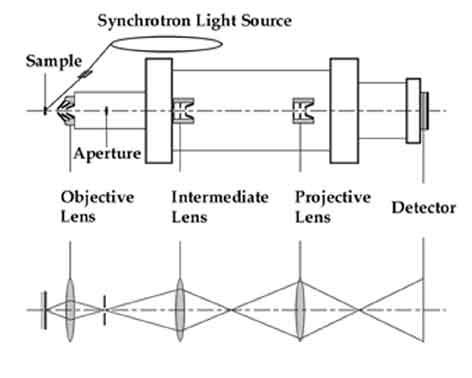 The MEPHISTO spectromicroscope uses three electrostatic lenses to form a photoelectron image at the detector (see figure). The sample is held at -10 to -20 kV while the first element is grounded, resulting in a strongly accelerating electric field between the sample and the optics. Each lens has three components, two grounded and the inner one held at -10 to -15 kV. The detector is a multichannel plate to ampify the electron signal, followed by a phosphor screen to provide the light image that is recorded by a video camera.
SPHINX
The SPHINX spectromicroscope uses six magnetic lenses, as well as stigmators to form a photoelectron image at the detector. The sample is held at -20 kV, while the first element is grounded, resulting in a strongly accelerating electric field between the sample and the optics. The detector is a multichannel plate that amplifies the electron signal, followed by a phosphor screen that provides the light image that is recorded by a video camera. The SPHINX achieves a better performance than MEPHISTO (1.5-fold higher electron transmission, and 5.5 nm resolution).
PEEM-3
For a description of the PEEM-3 microscope, see here.