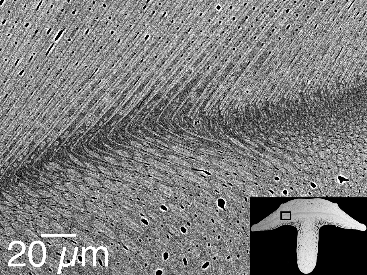
The teeth of sea urchins are among the most complicated structures in the natural world. The image here shows the cross section of one tooth which looks a bit like a mushroom. In the higher magnification image, you can appreciate the details of imbricated minerals: the elongated structures are plates (the rounded ones are fibers) and these both are entirely made of calcite. The space between fibers and plates is completely filled by high-Mg calcite nanoparticles ten nanometers in size.
- C. E. Killian, R. A. Metzler, Y. T. Gong, T. H. Churchill, I. C. Olson, V. Trubetskoy, M. B. Christensen, J. H. Fournelle, F. De Carlo, S. Cohen, J. Mahamid, A. Scholl, A. Young, A. Doran, F. H. Wilt, S. N. Coppersmith, and P.U.P.A. Gilbert.
Self-sharpening mechanism of the sea urchin tooth.
Adv. Funct. Mater. 21, 682-690 (2011). PDF - C. E. Killian, R. A. Metzler, Y. U. T. Gong, I. C. Olson, J. Aizenberg, Y. Politi, F. H. Wilt, A. Scholl, A. Young, A. Doran, M. Kunz, N. Tamura, S. N. Coppersmith, and P.U.P.A. Gilbert.
Mechanism of calcite co-orientation in the sea urchin tooth.
J. Am. Chem. Soc., 131, 18404-18409 (2009). PDF - Y. Ma, B. Aichmayer, O. Paris, P. Fratzl, A. Meibom, R. A. Metzler, Y. Politi, L. Addadi, P.U.P.A. Gilbert and S. Weiner.
The grinding tip of the sea urchin tooth: exquisite control over calcite crystal orientation and Mg distribution.
Procs. Natl. Acad. Sci. USA 106, 6048-6053 (2009). PDF SI New York Times article on this work - L Yang, CE Killian, M Kunz, N Tamura, and PUPA Gilbert.
Biomineral nanoparticles are space-filling.
R. Soc. Chem. – Nanoscale 3, 603-609 (2011).PDF - CE Killian, PUPA Gilbert.
Biomineral Single Crystals
Science 339, 510-511
Special Feature - AJ Lew, E Beniash, PUPA Gilbert*, MJ Buehler*.
Role of the mineral in the self-healing of cracks in human enamel.
DOI: 10.1021/acsnano.1c10407
ACS Nano 16, 10273–10280 (2022).