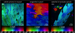
Polarization-dependent Imaging Contrast (PIC) mapping is a method introduced by Gilbert and her group to display the orientation of the crystaline c-axes carbonate biominerals.
PIC-mapping has been steadily improving since it was first introduced it in 2007(1), and is now fully quantitative in 2D(1-10), and most recently in 3D(11-13).
- RA Metzler, M Abrecht, RM Olabisi, D Ariosa, CJ Johnson, BH Frazer, SN Coppersmith, PUPA Gilbert.
Architecture of columnar nacre, and implications for its formation mechanism.
Phys. Rev. Lett. 98, 268102, 2007. - PUPA Gilbert, RA Metzler, D Zhou, A Scholl, A Doran, A Young, M Kunz, N Tamura, SN Coppersmith.
Gradual Ordering in Red Abalone Nacre.
J Am Chem Soc 130, 17519, 2008. - CE Killian, RA Metzler, YT Gong, IC Olson, J Aizenberg, Y Politi, FH Wilt, A Scholl, A Young, A Doran, M Kunz, N Tamura, SN Coppersmith, PUPA Gilbert.
The mechanism of calcite co-orientation in the sea urchin tooth.
J Am Chem Soc 131, 18404, 2009. - PUPA Gilbert, A Young, SN Coppersmith.
Measurement of c-axis angular orientation in calcite (CaCO3) nanocrystals using x-ray absorption spectroscopy.
Proc Natl Acad Sci USA 108, 11350, 2011. - CE Killian, RA Metzler, YUT Gong, TH Churchill, IC Olson, V Trubetskoy, MB Christensen, JH Fournelle, F De Carlo, S Cohen, J Mahamid, FH Wilt, A Scholl, A Young, A Doran, SN Coppersmith, PUPA Gilbert.
Self-sharpening mechanism of the sea urchin tooth.
Adv Funct Mater 21, 682, 2011. - PUPA Gilbert.
Polarization-dependent Imaging Contrast (PIC) mapping reveals nanocrystal orientation patterns in carbonate biominerals.
J Electr Spectrosc Rel Phenom, special issue on Photoelectron microscopy, Time-resolved pump-probe PES 185, 395, 2012. - IC Olson, R Kozdon, JW Valley, PUPA Gilbert.
Mollusk shell nacre ultrastructure correlates with environmental temperature and pressure.
J Am Chem Soc 134, 7351−7358, 2012. - IC Olson, RA Metzler, N Tamura, M Kunz, CE Killian, PUPA Gilbert.
Crystal lattice tilting in prismatic calcite.
J Struct Biol 183, 180, 2013. - IC Olson, AZ Blonsky, N Tamura, M Kunz, PUPA Gilbert.
Crystal nucleation and near-epitaxial growth in nacre.
J Struct Biol 184, 454, 2013. JOURNAL COVER. - PUPA Gilbert Photoemission spectromicroscopy for the biomineralogist In Biomineralization Sourcebook, Characterization of Biominerals and Biomimetic Materials; E DiMasi, LB Gower, Eds.; CRC Press: Boca Raton, FL, 2014, p 135.
- RT DeVol, RA Metzler, L Kabalah-Amitai, B Pokroy, Y Politi, A Gal, L Addadi, S Weiner, A Fernandez-Martinez, R Demichelis, JD Gale, J Ihli, FC Meldrum, AZ Blonsky, CE Killian, CB Salling, AT Young, MA Marcus, A Scholl, A Doran, C Jenkins, HA Bechtel, PUPA Gilbert.
Oxygen spectroscopy and Polarization-dependent Imaging Contrast (PIC)-mapping of calcium carbonate minerals and biominerals.
J Phys Chem B 118, 8449, 2014. - RT DeVol, C-Y Sun, MA Marcus, SN Coppersmith, SCB Myneni, PUPA Gilbert.
Nanoscale Transforming Mineral Phases in Fresh Nacre.
J Am Chem Soc 137, 13325, 2015. - B Pokroy, L Kabalah-Amitai, I Polishchuk, RT DeVol, AZ Blonsky, C-Y Sun, MA Marcus, A Scholl, PUPA Gilbert.
Narrowly distributed crystal orientation in biomineral vaterite.
Chem Mater 27, 6516, 2015.